The majority of lateral uka patients were female and had lower bmi than medial uka patients. As such, the vertical ligament found along the inside of the knee that attaches to this bony protrusion of the femur is known as the medial collateral ligament … The abaxial parts of the condyles are not articular and these rough area are. The medial and lateral condyles form the proximal part of the body of femur, and articulate with the proximal part of tibia to form the femorotibial joint. Physiotherapy is very important during the rehabilitation.

Medial in anatomy means toward the midline of the body, as opposed to lateral, or toward the sides of the body.
While both bones feature a medial and lateral condyle, with the lateral condyle on the other side of the knee, the medial condyle is the larger prominence because more weight is transferred across the inside aspect of the knee joint. Both condyles are smooth and rounded posteriorly to allow for motion and level out inferiorly allowing for articulation with the tibia. Physiotherapy is very important during the rehabilitation. The medial and lateral condyles form the proximal part of the body of femur, and articulate with the proximal part of tibia to form the femorotibial joint. Mar 14, 2022 · the medial femoral condyles are also distinguished by another anatomical term of location. They are separated by the deep intercondylar fossa, proximally bounded by the horizontal intercondylar line. Which femoral condyle is wider transversely? As such, the vertical ligament found along the inside of the knee that attaches to this bony protrusion of the femur is known as the medial collateral ligament … Feb 09, 2020 · the medial condyle is larger than the lateral (outer) condyle due to more weight bearing caused by the centre of mass being medial to the knee. Apr 06, 2022 · the medial and lateral condyles are the epiphyseal ends of the femur that articulate with the tibia and the patella. There is a significant difference in articular cartilage. If there is a fracture (break) in part of the condyle, this is known as a fracture of the femoral condyle. There are two condyles on each leg known as the medial and lateral femoral condyles.
The medial and lateral condyles form the proximal part of the body of femur, and articulate with the proximal part of tibia to form the femorotibial joint. There is a significant difference in articular cartilage. As such, the vertical ligament found along the inside of the knee that attaches to this bony protrusion of the femur is known as the medial collateral ligament … They are separated by the deep intercondylar fossa, proximally bounded by the horizontal intercondylar line. If there is a fracture (break) in part of the condyle, this is known as a fracture of the femoral condyle.

There are two condyles on each leg known as the medial and lateral femoral condyles.
Medial in anatomy means toward the midline of the body, as opposed to lateral, or toward the sides of the body. Feb 09, 2020 · the medial condyle is larger than the lateral (outer) condyle due to more weight bearing caused by the centre of mass being medial to the knee. Apr 06, 2022 · the medial and lateral condyles are the epiphyseal ends of the femur that articulate with the tibia and the patella. If there is a fracture (break) in part of the condyle, this is known as a fracture of the femoral condyle. Mar 14, 2022 · the medial femoral condyles are also distinguished by another anatomical term of location. The abaxial parts of the condyles are not articular and these rough area are. If there is a fracture (break) in part of the condyle, this is known as a fracture of the femoral condyle. The medial and lateral condyles form the proximal part of the body of femur, and articulate with the proximal part of tibia to form the femorotibial joint. Which femoral condyle is wider transversely? The majority of lateral uka patients were female and had lower bmi than medial uka patients. There are two condyles on each leg known as the medial and lateral femoral condyles. The medial and lateral condyles form the proximal part of the body of femur, and articulate with the proximal part of tibia to form the femorotibial joint. They are separated by the deep intercondylar fossa, proximally bounded by the horizontal intercondylar line.
As such, the vertical ligament found along the inside of the knee that attaches to this bony protrusion of the femur is known as the medial collateral ligament … If there is a fracture (break) in part of the condyle, this is known as a fracture of the femoral condyle. They are separated by the deep intercondylar fossa, proximally bounded by the horizontal intercondylar line. Feb 09, 2020 · the medial condyle is larger than the lateral (outer) condyle due to more weight bearing caused by the centre of mass being medial to the knee. Mar 14, 2022 · the medial femoral condyles are also distinguished by another anatomical term of location.
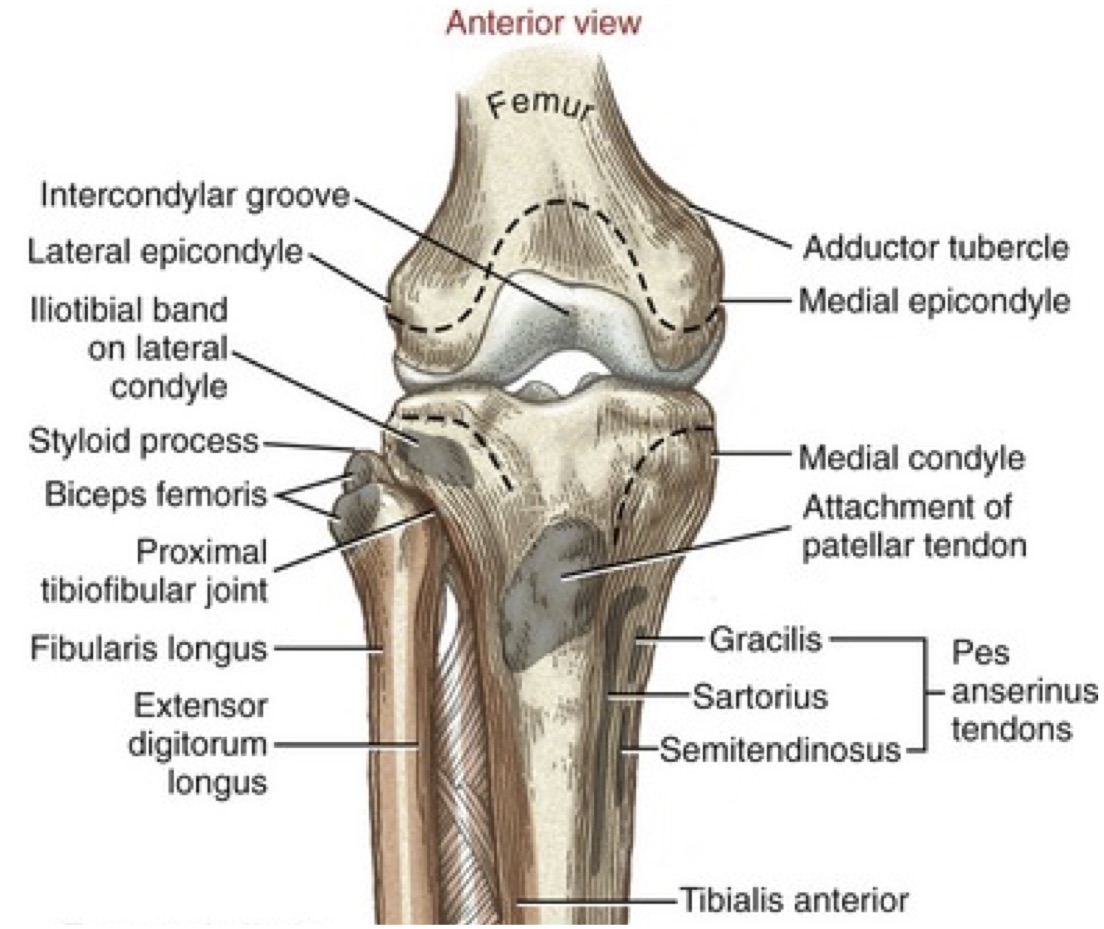
Feb 28, 2022 · a diagram of the knee, showing the medial condyles, where the medial collateral ligament attaches to the femur and tibia.
While both bones feature a medial and lateral condyle, with the lateral condyle on the other side of the knee, the medial condyle is the larger prominence because more weight is transferred across the inside aspect of the knee joint. Mar 14, 2022 · the medial femoral condyles are also distinguished by another anatomical term of location. Feb 09, 2020 · the medial condyle is larger than the lateral (outer) condyle due to more weight bearing caused by the centre of mass being medial to the knee. As such, the vertical ligament found along the inside of the knee that attaches to this bony protrusion of the femur is known as the medial collateral ligament … The majority of lateral uka patients were female and had lower bmi than medial uka patients. The abaxial parts of the condyles are not articular and these rough area are. If there is a fracture (break) in part of the condyle, this is known as a fracture of the femoral condyle. Apr 06, 2022 · the medial and lateral condyles are the epiphyseal ends of the femur that articulate with the tibia and the patella. There is a significant difference in articular cartilage. They are separated by the deep intercondylar fossa, proximally bounded by the horizontal intercondylar line. Medial in anatomy means toward the midline of the body, as opposed to lateral, or toward the sides of the body. They are separated by the deep intercondylar fossa, proximally bounded by the horizontal intercondylar line. Physiotherapy is very important during the rehabilitation.
Medial And Lateral Condyles Of Femur - Treatment Of Osteochondral Fracture Of The Lateral Femoral Condyle With Twinfix Ti Suture Anchor X Shaped Internal Fixation Under Arthroscopy A Surgical Technique And Three Cases Report Zhou 2020 Orthopaedic / They are separated by the deep intercondylar fossa, proximally bounded by the horizontal intercondylar line.. If there is a fracture (break) in part of the condyle, this is known as a fracture of the femoral condyle. There is a significant difference in articular cartilage. Feb 28, 2022 · a diagram of the knee, showing the medial condyles, where the medial collateral ligament attaches to the femur and tibia. Medial in anatomy means toward the midline of the body, as opposed to lateral, or toward the sides of the body. The medial and lateral condyles form the proximal part of the body of femur, and articulate with the proximal part of tibia to form the femorotibial joint.
0 comments
Post a Comment